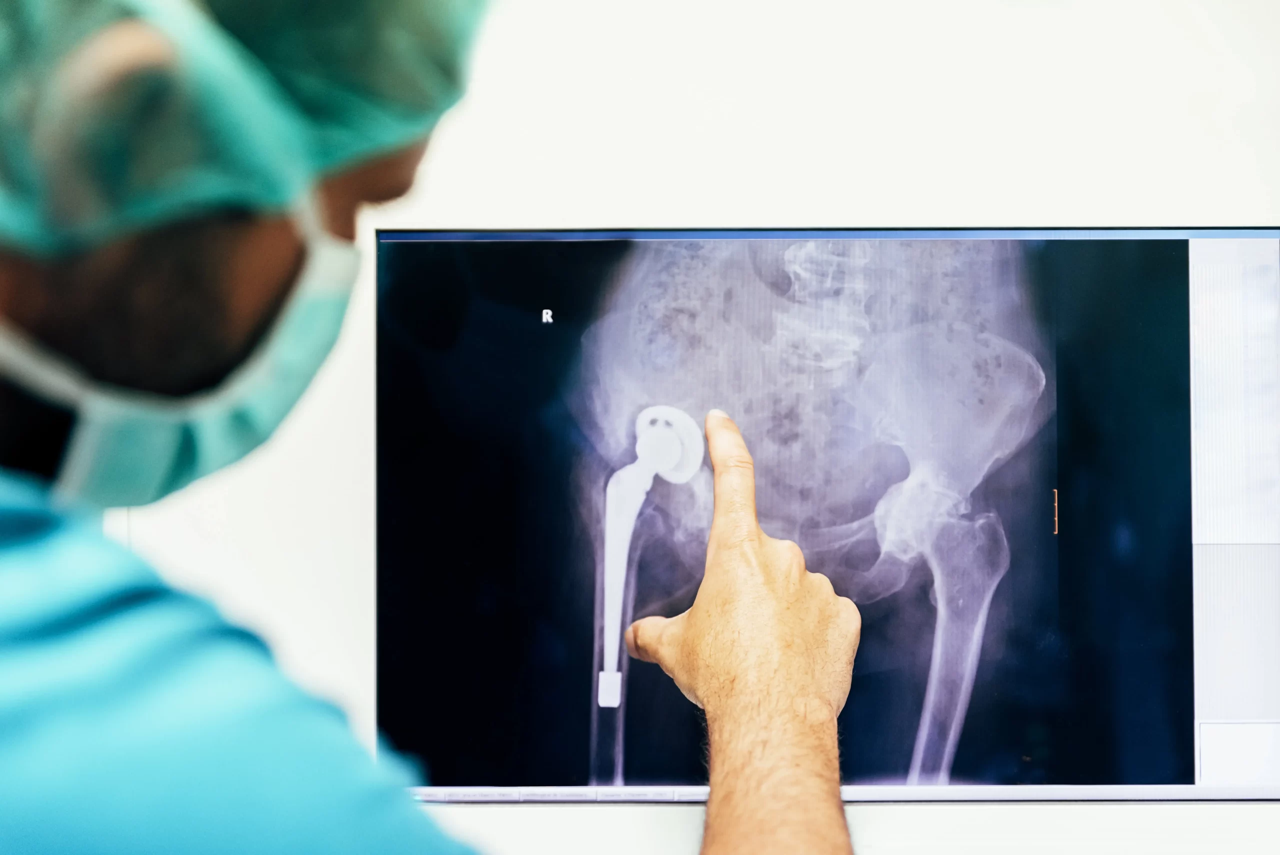
Preventive Hip Surgery And Its Goals
Preventive hip surgery means taking the necessary precautions in time and preventing the hip from becoming completely calcified and protecting it from a prosthesis surgery.

It is possible to use preventive hip surgery in every age group from the childhood to the adulthood. On the other hand, although not yet certain, there is an age limit of 45 years. Scientific publications on this subject indicate that the likelihood of success in the preventive hip surgeries performed after the age of 45 is reduced. However, it should be kept in mind that sometimes an individual at the age of 50 may have a bone quality at 40 years of age, whereas an individual at the age of 40 may have a bone quality at 50 years of age. Therefore, the age limit of 45 is a relative situation. Nevertheless, the chance of success increases when applied to the individuals under 45 years of age. It is not recommended after the age of 60.
Indications For The Preventive Hip Surgery
The hip joint is a ball-and-socket joint. The head of the bone is a rounded ball and the socket has a hollow shape corresponding to this ball and operates with zero error. In some cases, the fact that the socket is deeper than the normal situation or completely rounded, and that the head has a ridge or protrusion in any part thereof causes the hip to rub in use over the years, thereby leading wear in these regions. In addition, if the bone is not fully rounded, that is, if there is a structural problem from the childhood, no sign is seen as the cartilage is initially too thick, whereas when the adult age is reached after years, pain occurs and walking problems may be seen due to the friction of the hip. In such cases, preventive hip surgery is implemented as an early treatment of the problems caused by an unconformity of the socket.
Complaints in Patients
Usually, a hip pain occurs in the patients. Pain is described by the patients as an inguinal pain that spreads towards the knee. Since the pain is in the inguinal region, women tend to consult primarily to the medicinal branches such as gynecology and urology. In some cases, the patients may consult doctor with the complaint of low back pain. Firstly, a differential diagnosis should be made as the low back pain may be confused with gynecological diseases and urinary tract infections. A radiological examination should be performed to make the diagnosis. The diagnosis at a level of 90% may be made by a plain radiograph taken in different positions, for which the measurements are performed. MRI, three-dimensional tomography and the other tests may be needed while the treatment is planned. However, it should not be thought that every radiological problem will be treated. There should be a waiting period, and then it should be controlled whether the patient complaints about pain. In some cases, although there is a radiological problem in the individuals of 70-80 years of age, it is seen that no complete calcification and no complaint about pain is present. However, in the case that pain is present and the diagnosis is made, it is necessary to start treatment immediately in order to prevent calcification of the hip completely.
Preventive Hip Surgeries and Hip Arthroscopy
There are two types of treatment, such as open and closed surgery, for the problems such as hip impingement or incongruence. Open surgery is also divided into major-open and mini-open surgery. As it can be understood from the name, the open surgery is applied by opening and reaching the hip and treating the problem. Sometimes, if the problem is smaller and localized to a certain area, the treatment is performed via a small incision of five to six centimeters and this is called “mini open”.
The application of arthroscopy, called the closed surgery, has gained momentum, especially over the last decade. It can be applied for both children and adults. Hip arthroscopy is performed by imaging the hip with a camera and the other auxiliary tools, which are inserted through small incisions, and treating the joint-related diseases. Nowadays, it can be applied in almost every hip-related problem but there are limitations. Unlike the other joints, special angle cameras are used, as the hip is at a deep level and the space for the insertion and movement of the tools is very small. Thus, the entire hip may be substantially imaged. However, in the case that the hip is too deep and calcified, the camera cannot be inserted through the spaces between the joints. Besides this, it is also possible to use arthroscopy at a rate of 90%.
Contraindications for Arthroscopy
When it is thought that there is an active infection in the hip, care should be taken in this group. If the hip is in a condition called “adhered”, i.e. if it seems that an adhesive is applied therein, no opening process may be performed in the hip and insertion of a camera may be detrimental. In these cases, arthroscopy is not performed as no force should be applied.
On the other hand, a special rehabilitation program is required after arthroscopy. Patients are warned about movements that should not be done. The length of hospital stay is usually one day. If the person being treated is an active athlete, he/she can continue playing sport after 3 months. In some cases, this period may increase up to 6 months.

